Why are large imaging files a growing challenge in research labs at year-end?
As research institutes and life science laboratories face the year-end rush, one often overlooked hurdle is the surge in imaging data. Modern tools generate extremely large files – sometimes gigabytes per sample. Multiplied across hundreds or thousands of datasets, this can strain storage systems and IT workflows, risking delays when deadlines are most critical.
The impact of overwhelming data loads
Without the right tools, labs may face storage capacity issues, slow rendering, and longer wait times to review datasets. This reduces throughput and jeopardises the timely completion of experiments, publications, and grant reports – especially during peak year-end periods.
Smarter image analysis with Agilent BioTek Gen5 Software
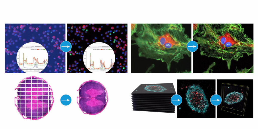
Agilent BioTek Gen5 Software for Imaging & Microscopy streamlines image capture, processing, and analysis within one interface. Researchers can design 2D or 3D workflows in widefield or confocal imaging, supported by modules for in-depth analysis across a wide range of cell applications.
Key capabilities include:
Single image capture, montaging, Z-stacking, and beaconing
Advanced processing such as background removal, deconvolution, stitching, Z-projection, and 3D rendering
Automated functions including confluence determination, object counting, neurite outgrowth analysis, spot counting, and single object tracking
Built-in tools for exporting high-resolution images, graphs, and kinetic movies
By reducing technical bottlenecks, Gen5 helps researchers generate insights faster – a real advantage when deadlines are tight.
Biofilms have an inherent three-dimensional structure that can be reconstructed using confocal microscopy. While widefield imaging captures compositional properties, confocal laser scanning microscopy (CLSM) reduces out-of-focus light and background interference, improving image quality when analysing live biofilms at varying focal depths.
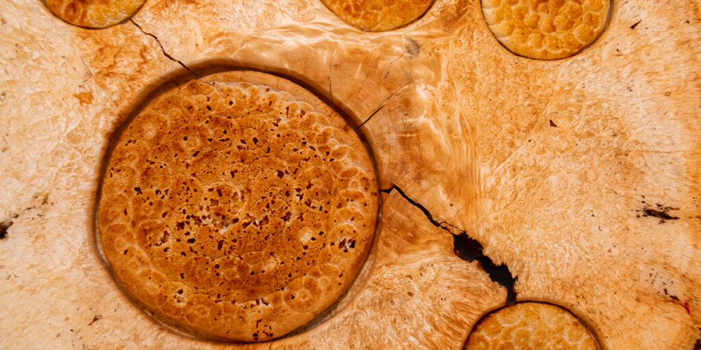
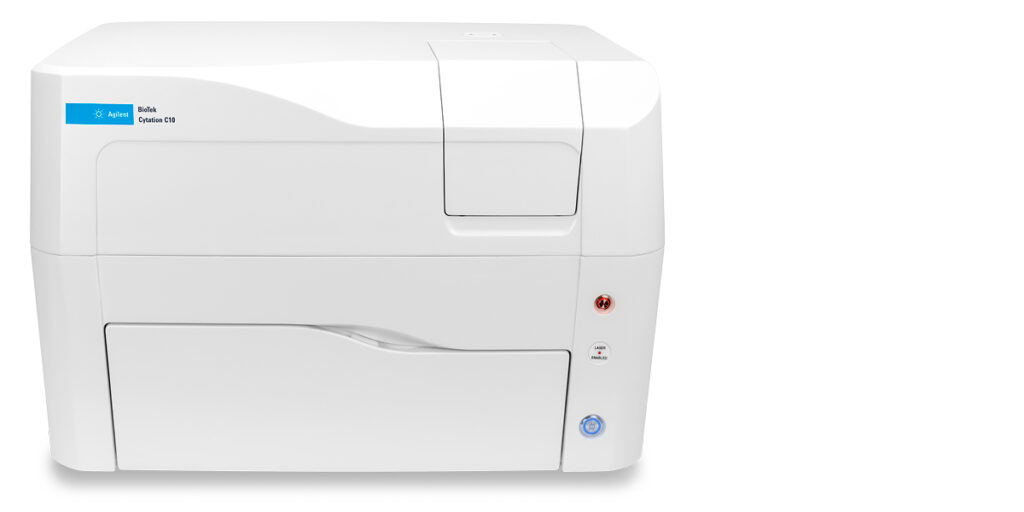
In addition to managing large image files, life science researchers are also exploring deeper biological insights – such as how microorganisms form biofilms on tissues, experimental surfaces, or model systems, which can influence disease models and basic biological research. Agilent’s solutions underscore the importance of properly visualising these complex, three-dimensional microbial communities. The Cytation C10 Confocal Imaging Reader is well-suited for biofilm research. Combining widefield and confocal modes with environmental control, it captures high-quality, live-cell images of thick multicellular structures. Confocal optics layered over plate-reader modalities allow researchers to visualise biofilm architecture and metabolic activity while keeping file sizes manageable.
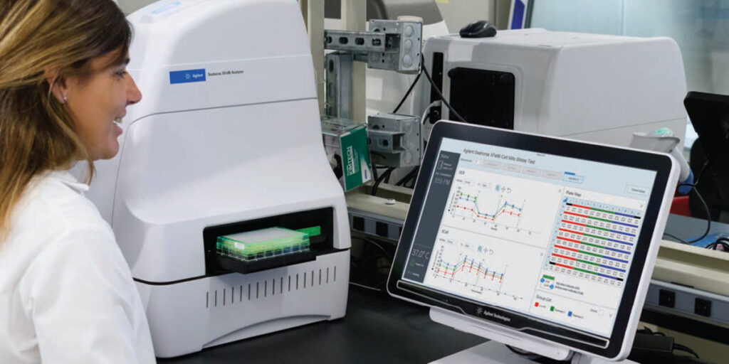
Researchers also need insights that go beyond imaging data. The new 
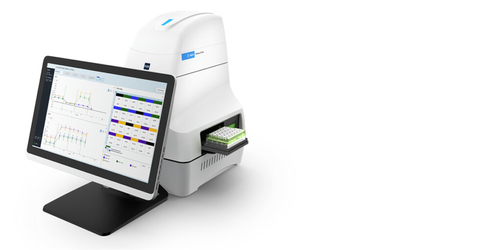
Agilent Seahorse XF Flex Analyzer complements imaging by enabling real-time analysis of live-cell metabolism in both 2D and 3D models. This provides functional insights without adding to data storage burdens, helping labs interpret results faster.
Automated workflows for file transfer, compression, and retrieval reduce manual workload and delays. This frees scientists to focus on data interpretation rather than technical troubleshooting, improving accuracy and turnaround times.
South African research institutions balance academic projects and industry collaborations, each with unique reporting demands. Scalable imaging systems help labs manage seasonal surges and diverse sample types without compromising quality or speed.
Ready to optimise your lab’s workflows this festive season?
Agilent’s advanced imaging solutions – including the Cytation C10 Confocal Imaging Reader, BioTek Gen5 Software, and Seahorse XF Flex Analyzer – help labs improve efficiency, reduce delays, and deliver results faster. Complementary methods like holotomography can provide additional insights, expanding analytical possibilities.
Contact us to learn more about Agilent’s solutions for life science research.
While cancer’s visible symptoms capture our attention, it’s the invisible metabolic processes within cells that truly drive the disease. Cancer cells reprogram their metabolism to fuel rapid growth, resist treatments, and evade the immune system. Understanding these metabolic changes isn’t just academic curiosity; it’s the key to developing more effective, targeted therapies that can outsmart cancer at its most fundamental level
The challenge: Decoding cancer’s metabolic complexity
Cancer metabolism is far more intricate than simply “cells growing fast.” Tumour cells undergo profound metabolic reprogramming, switching energy production pathways, altering nutrient consumption, and even changing how they communicate with surrounding tissues. This metabolic flexibility makes cancer particularly challenging to treat.
Traditional research methods often provide only snapshots of cellular activity, missing the dynamic changes that occur over time. What researchers need are technologies that can monitor these metabolic processes as they happen, providing real-time insights into how cancer cells behave and respond to potential treatments.
Modern cancer research demands technologies that can capture the full picture of cellular metabolism in real-time. Two revolutionary platforms are transforming how scientists study cancer metabolism, offering unprecedented insights into cellular behaviour and treatment responses.
Comprehensive metabolic profiling with Seahorse technology
Oxygen Consumption Rate(OCR) and Extracellular Acidification Rate(ECAR) are key indicators of mitochondrial respiration and glycolysis and these measurements provide a systems-level view of cellular metabolic function in cultured cells. The Agilent Seahorse XFe96 Analyzer represents a breakthrough in metabolic analysis, allowing researchers to measure both OCR and ECAR simultaneously in live cells.
Key capabilities for cancer research:
Real-Time Metabolic Measurements: Monitor how cancer cells switch between different energy production pathways, revealing metabolic vulnerabilities that could be targeted therapeutically.
Drug Response Analysis: Agilent Seahorse XF technology provides critical functional measurements in live cells to identify potential druggable gene and protein targets, and to validate their role in cancer cell proliferation, adaptation, and survival.
High-Throughput Screening: The 96-well format enables researchers to test multiple conditions simultaneously, accelerating the pace of discovery in metabolic drug development.
Dynamic cell behaviour analysis with xCELLigence technology
The Agilent xCELLigence RTCA eSight is an advanced real-time cell analysis system that enables comprehensive insight into a cell’s health, behaviour, function, and biological processes using live, simultaneous, and real-time biosensor-based and image-based measurements.
This innovative platform combines impedance-based monitoring with live-cell imaging, providing a complete picture of cellular responses to metabolic interventions.
Key advantages for cancer metabolism studies:
Label-Free Monitoring: The xCELLigence RTCA instrument uses noninvasive electrical impedance monitoring to quantify cell proliferation, morphology change, and attachment quality in a label-free, real-time manner, allowing for continuous observation without disturbing cellular processes.
Multi-Modal Analysis: Simultaneous impedance measurements and fluorescence imaging provide complementary data on cell viability, proliferation, and metabolic activity.
Treatment Response Tracking: Monitor how cancer cells respond to metabolic inhibitors or combination therapies over extended periods, revealing both immediate and delayed effects.
Morphological Changes: Observe real-time changes in cell morphology that accompany metabolic reprogramming, providing visual confirmation of cellular responses.
By combining these advanced technologies, researchers can now:
Identify Metabolic Vulnerabilities: Discover specific metabolic pathways that cancer cells depend on for survival, revealing new therapeutic targets.
Optimise Combination Therapies: Understand how metabolic inhibitors work in combination with traditional treatments, potentially improving efficacy while reducing side effects.
Personalise Treatment Approaches: Characterise the metabolic profiles of different cancer types and subtypes, enabling more precise treatment selection.
Accelerate Drug Development: Rapidly screen potential metabolic inhibitors and validate their mechanisms of action in relevant cellular models.
Your gateway to advanced cancer metabolism research
The future of cancer treatment lies in understanding and targeting the metabolic engines that drive tumour growth and resistance. Chemetrix provides researchers with access to these cutting-edge technologies, along with the expertise needed to unlock their full potential.
Our comprehensive support includes:
Expert consultation on experimental design and methodology
Technical training and ongoing support
Method development and optimisation
Data analysis and interpretation guidance
Ready to advance your cancer metabolism research?
The battle against cancer requires the most sophisticated tools science can provide. With Seahorse and xCELLigence technologies, you have the power to see cancer metabolism in real-time, revealing new possibilities for treatment and hope for patients.
Cell culture lies at the heart of many biological studies, serving as a foundational technique for a myriad of applications, from basic research to drug discovery. Register for this exciting webinar that will explore the newest features and instrument capabilities supporting kinetic live-cell imaging, from real-time analysis of rapid GPCR signalling pathways to long-term treatment-induced effects on cell proliferation and viability.
Speaker
Joe Clayton, PhD Scientific Program Manager, Agilent Cell Analysis
With over 20,000 genes, 200,000 proteins and thousands of pathways, you can’t measure everything in a cell at once, but you can measure the energy that drives them. Chemetrix offers cell analysis technology solutions that detect discrete changes in cellular bioenergetics in real-time. Real-time cell measurements provide a clear window into the critical functions driving cell signaling, proliferation, activation, toxicity and biosynthesis. Move beyond analysing what your cells are, and reveal a clearer measure of what they do.
Best Practices & Fundamentals
Procedures, Techniques and Resources for a Successful Xf Assay.
Training
Energy Pathways Video
Seahorse XF Training Webinar – Gather Assay Materials