Why are large imaging files a growing challenge in research labs at year-end?
As research institutes and life science laboratories face the year-end rush, one often overlooked hurdle is the surge in imaging data. Modern tools generate extremely large files – sometimes gigabytes per sample. Multiplied across hundreds or thousands of datasets, this can strain storage systems and IT workflows, risking delays when deadlines are most critical.
The impact of overwhelming data loads
Without the right tools, labs may face storage capacity issues, slow rendering, and longer wait times to review datasets. This reduces throughput and jeopardises the timely completion of experiments, publications, and grant reports – especially during peak year-end periods.

Smarter image analysis with Agilent BioTek Gen5 Software
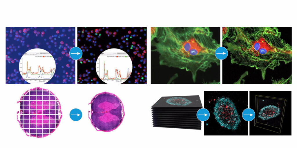
Agilent BioTek Gen5 Software for Imaging & Microscopy streamlines image capture, processing, and analysis within one interface. Researchers can design 2D or 3D workflows in widefield or confocal imaging, supported by modules for in-depth analysis across a wide range of cell applications.
Key capabilities include:
- Single image capture, montaging, Z-stacking, and beaconing
- Advanced processing such as background removal, deconvolution, stitching, Z-projection, and 3D rendering
- Automated functions including confluence determination, object counting, neurite outgrowth analysis, spot counting, and single object tracking
- Built-in tools for exporting high-resolution images, graphs, and kinetic movies
By reducing technical bottlenecks, Gen5 helps researchers generate insights faster – a real advantage when deadlines are tight.
Biofilm characterisation
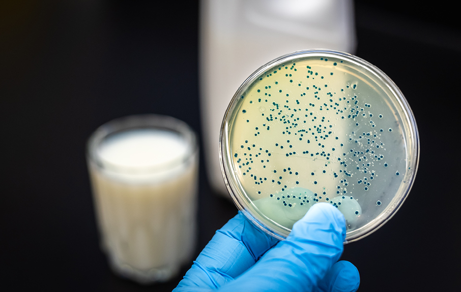
Biofilms have an inherent three-dimensional structure that can be reconstructed using confocal microscopy. While widefield imaging captures compositional properties, confocal laser scanning microscopy (CLSM) reduces out-of-focus light and background interference, improving image quality when analysing live biofilms at varying focal depths.

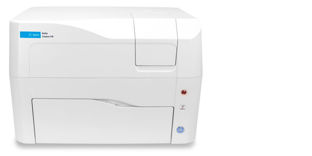
In addition to managing large image files, life science researchers are also exploring deeper biological insights – such as how microorganisms form biofilms on tissues, experimental surfaces, or model systems, which can influence disease models and basic biological research. Agilent’s solutions underscore the importance of properly visualising these complex, three-dimensional microbial communities. The Cytation C10 Confocal Imaging Reader is well-suited for biofilm research. Combining widefield and confocal modes with environmental control, it captures high-quality, live-cell images of thick multicellular structures. Confocal optics layered over plate-reader modalities allow researchers to visualise biofilm architecture and metabolic activity while keeping file sizes manageable.
Beyond imaging
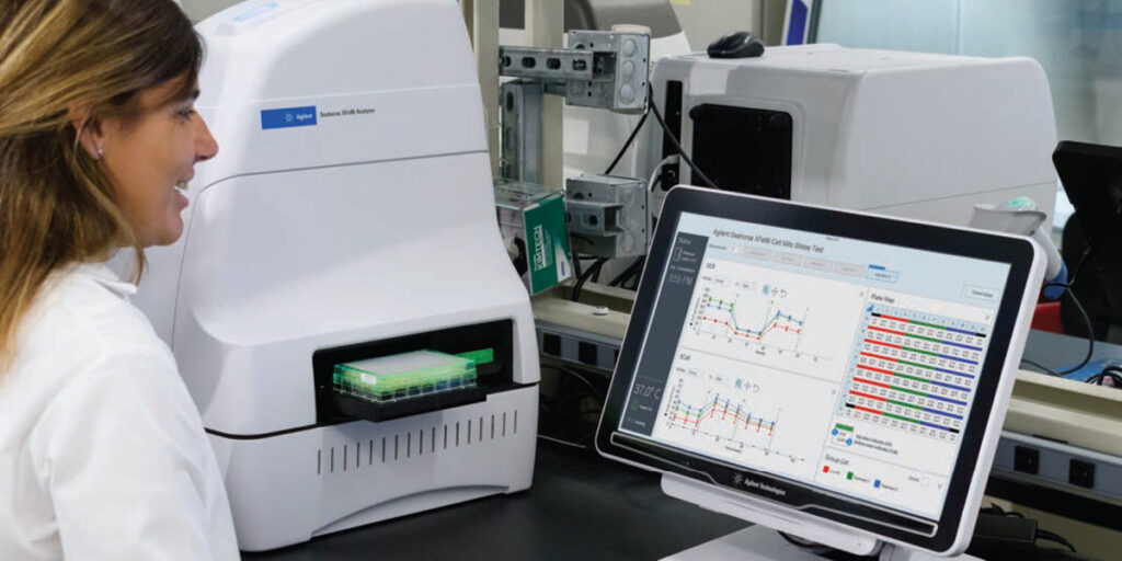
Researchers also need insights that go beyond imaging data. The new Agilent Seahorse XF Flex Analyzer complements imaging by enabling real-time analysis of live-cell metabolism in both 2D and 3D models. This provides functional insights without adding to data storage burdens, helping labs interpret results faster.
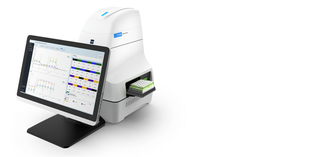
Agilent Seahorse XF Flex Analyzer
Why is automation matters
Automated workflows for file transfer, compression, and retrieval reduce manual workload and delays. This frees scientists to focus on data interpretation rather than technical troubleshooting, improving accuracy and turnaround times.
Why scalability is vital for South African labs
South African research institutions balance academic projects and industry collaborations, each with unique reporting demands. Scalable imaging systems help labs manage seasonal surges and diverse sample types without compromising quality or speed.
Ready to optimise your lab’s workflows this festive season?
Agilent’s advanced imaging solutions – including the Cytation C10 Confocal Imaging Reader, BioTek Gen5 Software, and Seahorse XF Flex Analyzer – help labs improve efficiency, reduce delays, and deliver results faster. Complementary methods like holotomography can provide additional insights, expanding analytical possibilities.
Contact us to learn more about Agilent’s solutions for life science research.

